Clinical and Radiographic Examination of the Equine Foot
- Nanric
- Nov 21, 2003
- 37 min read
Updated: Apr 21, 2020
49th Annual Convention of the American Association of Equine Practitioners, 2003, New Orleans, Louisiana
THE EQUINE FOOT, IN-DEPTH
Clinical and Radiographic Examination of the Equine Foot (21-Nov-2003)
R. F. Redden
Versailles, KY, USA.
1. Introduction
Lameness is one of the most frequently encountered problems in equine practice. The foot is involved, either directly or indirectly, in the large majority of lameness cases, as it is the first line of defense for the animal. The health of the foot plays a major role in the fight or flight response that has preserved this noble species for several thousand years. "No foot, no horse" is an adage that has been used across the world for centuries. This indisputable statement encapsulates the importance of a healthy foot; yet we know less about the foot than about almost any other part of the horse, and it is the one piece of anatomy that is dependent on a lay profession for the preservation of its health and function.
Worldwide, farriers bear much of the responsibility for maintaining or restoring the health of the horse's foot. For centuries their knowledge and skills have been self-taught, without the benefit of a formal educational program. Their basic job description is to keep the foot healthy by using effective but primitive methods to control the ill effects of horn growth and of wear and tear on the hoof capsule, with little or no information about the effects of these procedures on the sensitive soft tissues, vascular supply, or bone. Veterinarians, on the other hand, have been taught anatomy, physiology, and basic examination techniques; however, they often have limited working knowledge of the foot and little or no farriery skills.
Both professions play important and complementary roles. Veterinarians and farriers alike are often asked to examine the foot for a variety of reasons, including developmental problems, gait analysis, lameness exams, and prepurchase exams. In many cases, the opinions that result are as diverse as the backgrounds and areas of expertise of the respective professionals. Combining the knowledge and skills of a competent farrier with the medical and surgical training of the veterinarian greatly enhances the diagnostic and prognostic potential of both clinical and radiographic examinations. Working together also advances the professional standing of veterinarians and farriers.
Clinical and radiographic examinations of the foot are simply discovery exercises. Numerous authors have described their methods and techniques in detail. But despite the vast amount of written material on the subject, obtaining meaningful information about the foot remains a challenge for veterinarians and farriers. The key is to use a disciplined, methodical approach that is designed to disclose and define the various normal soft tissue parameters, normal bone anatomy, normal hoof capsule anatomy, and how each component is interrelated. The protocol should also reveal the response of these structures to the forces imposed by ground contact, supporting tissues, and the horse's body weight.
Seeking and defining specific pieces of information in a consistent, repeatable manner for each foot, in each horse, greatly enhances the practitioner's understanding and knowledge bank regarding the vast range of normal-which is the real information you want. Whether examining a foot or a radiograph, look for all the normal areas first; what's left over points to the problem you seek. This simple approach effectively helps avoid misinterpretation, a common result of forming an opinion without sufficient diagnostic information; for example, making presumptions concerning the clinical relevance of a radiographic lesion without consideration of the history or physical findings.
2. Clinical Examination
Regardless of the purpose of the examination, the physical exam is the most important aspect of evaluating the equine foot. The extent and nature of the exam must be tailored to the situation, however, taking into account the demands of the client. Good horsemanship, a good working knowledge of the foot, and some basic farriery skills are other prerequisites for a proper and safe examination.
A complete history which clearly describes the complaint complements the physical exam and adds context to any clinical findings. Listen to the history as you examine the foot, but do not jump to conclusions nor be swayed by the opinions or conclusions of others. Visually inspect the foot before picking it up, and feel the hoof capsule with your hands, noting its many unique characteristics.
Although certain generalities can be made, there is a range of normal for hoof characteristics which is influenced by the horse's breed, age, environment, and use. Considering the variability imposed by these factors, the range of normal can be very broad. The importance of understanding the variability in structure of the healthy equine foot lies in identifying subtle deviations from normal which are of clinical significance. These early distortions are easily missed if the normal parameters for a horse of that breed, age, environment, and use are not appreciated.
The following example details the requirements for adequately defining normal for a particular horse. Let us consider the forefoot of a 3-yr-old Thoroughbred horse, bred for racing but used as a noncompetitive riding horse in central Kentucky. That foot would probably have the following characteristics: a hoof angle between 50 degrees and 58 degrees, and a heel angle perhaps 15-20 degrees less; a relatively straight wall (i.e. no flaring, dishing, or bulging); width approximately 5 in. (measured at the widest point); mass of digital cushion 2-3 in. (discussed later); hoof wall thickness of 3/8-1/2 in. at the toe and the bars;a hoof wall perhaps one-half as thick at the quarters; a sole with a moderate cup (3-5 mm in height); a frog in contact with the ground (although it would also be normal for this horse to have a relatively flat sole, i.e., little or no cup, and a large, flat frog); and a hoof wall with a solid appearance and a glossy surface.
We must leave behind the "ideal" of the normal equine foot depicted by artists in veterinary and farriery texts for the past century or more. Hoof angles and heel angles do not match on any normal foot. And the "ideal" toe angles of 45 degrees for front feet and 50 degrees for hind feet are far from normal as they do not match the pastern angles. One must become a connoisseur of horses' feet and begin to build a personal data bank of normal for particular breeds, age groups, environments, and uses.
The Seat of Pain
When dealing with a lame horse, most authors consider the physical exam simply a means of reaching a diagnosis, i.e., of giving the problem a name. While this approach certainly satisfies one of the goals of the exam (to identify the problem), years of experience as an equine podiatrist have made me very aware that most owners want a fix and could care less about a diagnosis.
As I'm going through a lameness work-up, I focus on identifying the area(s) of pain rather than specific pathology. Dividing the foot into two halves, front and back, then dividing further into quadrants (medial and lateral, front and back) offers a simple way of isolating the specific area of inflammation or seat of pain (Fig. 1, A and B).
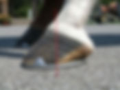
Figure 1. (A) Imagine dividing the foot in half.
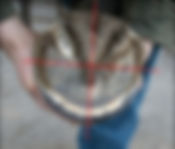
Figure 1. (B) Then in quarters.
Dividing the foot into four basic zones helps me determine whether the components in each zone fit within the range of normal for that particular animal. With my understanding of radiographic anatomy (again bearing in mind the range of normal), I visualize the bone and associated soft tissues superimposed over the hoof (Fig. 2). Any finding that falls outside the range of normal is considered relevant, as it contributes to the dysfunction of the foot as an integrated unit and thus probably plays a role in the current lameness problem.
Simply cleaning the ground surface of the hoof can reveal areas of possible concern. (Fig. 3). Each of these areas is a map of a potential problem: examine each thoroughly before moving on. Remember to look for all the normal areas first, and what is leftover often points to the problem that you are attempting to identify.

Figure 2. Visualize the bone and associated soft tissues
superimposed over the hoof.
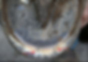
Figure 3. Simply cleaning the ground surface of the
hoof can reveal areas of possible concern.
After a quick visual exam, I palpate, using thumb pressure to locate areas of increased sensitivity along the coronary band, the bulbs of the heel, and even over the sole on thin-soled feet. Hoof testers should be used with great care, because inappropriate use causes the horse to anticipate further pain and show an exaggerated response to even light pressure.
When applying hoof testers, use a very soft touch. All that is needed to identify areas of increased sensitivity is just enough pressure to cause slight movement of thin horn (e.g., the sole in a thin-soled horse). Also be aware of how you are holding the horse's leg. If, by positioning the limb between your knees so that you are comfortable, the horse is made uncomfortable, you may elicit a response that has nothing to do with the foot.
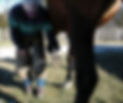
It is easy to abduct the limb too far when placing the horse's lower limb between your knees. To avoid this situation, note where the horse's body in relation to the foot when you first pick up the leg. Try to maintain that orientation when placing the limb between your knees-i.e., put yourself where the foot is or have someone hold the limb for you (Fig. 4 A-D).
4A 4B

4C 4D


Figure 4. (A) Note relaxed position of foot.
(B) Position yourself to horse’s relaxed position.
(C) Avoid abducting limb for your comfort.
(D) Proper stance when using hoof testers.
The Failing Structure
Distinguishing the abnormal area(s) allows me to identify which part(s) of the system is failing and affecting the integrity of the whole. Simply recognizing the failing structure(s) as the primary problem-the underlying cause of any secondary bone and/or soft tissue disease-gives new meaning to the discovery exercise and places new emphasis on the findings. Following is an example of this concept. Race horses, or in fact any speed horse, with less than 10 mm of sole, zero or negative palmar angle (the angle of the palmar margin of PIII relative to the ground surface), loss of cushion mass (see below), obvious medial-lateral imbalance, and a history of foot pain are often diagnosed with navicular disease, pedal osteitis, or bruised feet. Any of these diagnoses may be correct and the associated pathology may be contributing to the present lameness. However, more important is the fact that the essential protective function of the hoof capsule and the shock-absorbing features of the cushion network are seriously compromised, and the cumulative effects of these failing systems are now of paramount importance.
The "diagnosis" in this case is thus, multifaceted. However, it can be simplified by describing the situation as one of mild, moderate, or excessive horn loss associated with mild, moderate, or excessive compromise of the soft tissues. Instead of being focused on a medical diagnosis (which may well be challenged by another veterinarian or farrier) and a quick fix to satisfy the immediate demands of the client, identifying the failing systems allows the focus to be placed on a solution, which in this case involves restoring the much-needed hoof mass.

Figure 5. Use thumb and finger to guesstimate depth of digital cushion.
The depth of the digital cushion can be estimated by placing your thumb in the shallow depression between the heel bulbs and placing the index finger of the same hand on the center of the frog (Fig. 5). In light breed horses with strong, healthy heels, the distance between thumb and fingertip is in the range of 3-3.5 inches.
6A 6B
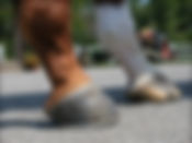

Figure 6. (A) Typical Thoroughbred hind foot. Note coronary band relationship with the ground. (B) Front foot, American Saddlebred. Growth ring patterns, coronary
band conformation, heel tubule angles, toe angles, and horn quality offer insight to sole depth, palmar angle, and overall state of balance.
When this distance is well short of the normal range, one can expect to see evidence of soft tissue compromise radiographically. This simple observation, coupled with noting the slope of the coronary band relative to the ground, also allows an estimation of sole depth and palmar angle.
Figures 6 and 7 illustrate how these observations correlate with radiographic findings. Note the difference in slope of the coronary band, angle of the horn tubules at the heel, and depth of cushion between the two horses (Fig. 6A, 6B). Compare these photographs with lateral radiographs of the same feet (Fig. 7A, 7B).
7A 7B

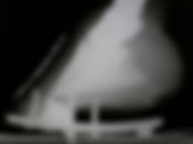
Figure 7. Radiographs of feet shown in Figure 6. (A) Note negative 6 degree palmar angle. (B) Note H-L zone and positive 6 degree palmar angle.
Incidentally, in my experience hind feet with a zero or negative plantar angle (wings of PIII level with or lower than the apex) are often associated with pain in the lumbar area or croup. Back pain in these horses frequently diminishes once heel mass is improved and a normal plantar angle is restored.
Other Notes
It is necessary to remove the horse's shoe in order to thoroughly examine the foot, especially when the primary problem cannot be identified with the shoe on. At the very least, the shoe prevents examination of the bearing surface of the wall, the terminal laminae, and the perimeter of the sole. However, care must be taken when pulling shoes. In feet with fragile walls, raised nail clinches, or a special shoeing package, the shoe is best removed by a competent farrier unless you have considerable farriery expertise.
A good sense of smell can be a valuable aid in examining the foot. We all know the smell of a foot with thrush. But your olfactory sense can also help you identify digital sepsis. With experience, it is even possible to distinguish soft tissue necrosis from septic processes involving bone. Again, attention to detail is the key to refining one's examination skills.
Key Points
physical examination is the most important part of evaluating the foot
develop a methodical approach, and use it every time
look for normal first (bearing in mind the range of normal for that horse's breed, age, environment, and use); what's left over points to the problem you seek
localize the seat of pain to one or more quadrants
visualize the underlying bone and associated soft tissues when looking at the hoof
think in terms of identifying the failing structure(s)
3. Radiographic Examination Much has been written about specific views for imaging the equine foot. Almost without exception, the primary objective of these views is examination of bone (PIII, navicular bone, and/or coffin joint surfaces). Little or no attention is paid to the soft tissues within the hoof capsule. This approach seriously limits the scope and accuracy of the radiographic examination and thus its value in developing an action plan for managing lameness involving the foot.
The coffin bone is suspended within its protective shell by soft tissues whose health is crucial to the structural and functional integrity of the foot as a whole. Dysfunction is inevitable when any of the soft tissues are compromised or strained beyond their normal limits. Over the many years I have worked as an equine podiatrist, I've come to appreciate the fact that soft tissue pathology is present to some degree in every footsore horse. Thus, evaluation of the soft tissue zones within the hoof capsule is an extremely important part of radiographic examination of the foot.
Evaluating the Soft Tissues
While it is true that radiography is relatively poor at imaging soft tissues, a lot of information about the soft tissues within the foot can be gleaned from good quality radiographs taken with soft tissue detail in mind (discussed in the next section). At the very least, the width of the corium and horn can be accurately measured for both hoof wall and sole, provided the outer surface of the dorsal hoof wall is delineated using radiopaque material and the ground surface is defined either by the shoe or by a radiopaque marker in the surface of the positioning block. I measure the following indices on all routine lateral films (Fig. 8).
Sole Depth
Sole depth is defined as the vertical distance between the palmar/plantar margin of PIII and the outer surface of the sole. It is routinely measured at the distal tip, or apex, of PIII (Fig. 8). A normal, healthy foot has a sole depth of at least 15 mm. Based on venographic studies in a wide variety of horses, I consider a sole depth of less than 15 mm to be clinically significant. In a normal foot, the papillae of the solar corium appear to need a space of at least 10 mm between the palmar surface of PIII and the cornified layer of the sole for adequate vascular filling; and at least 5 mm of cornified sole is required to protect the solar corium. Venograms in horses with a sole depth <15 mm show solar papillae that are bent, compressed, or even absent. This distortion or compression surely inhibits sole growth, creating a vicious cycle of thin, tender soles.

Figure 8. Standard low beam, soft tissue view with opaque wall marker and ground surface marker offers a consistent means of accurately measuring soft tissue parameters. Progressive farriers often use this view as a blueprint for pathological shoeing.
Dorsal Horn-Lamellar Zone Width
Dorsal horn-lamellar (H-L) zone width is defined as the distance between the dorsal surface of PIII and the outer surface of the dorsal hoof wall, measured with the ruler perpendicular to the dorsal surface of PIII (Fig. 8). Dorsal H-L zone width can be measured anywhere along the dorsal face of PIII, but I routinely measure it at two locations: just below the extensor process, and near the distal tip of PIII. I record the measurements as proximal/distal (e.g. 15/15, meaning that the dorsal H-L zone is 15 mm at both locations). In a normal adult foot, the measurements should be the same proximally as distally (i.e. both numbers are identical). In the immature foot, the proximal value may be greater than the distal value.
Normal dorsal H-L zone width in Quarter Horses, Thoroughbreds, and most other light horse breeds is 15-16 mm. In Standardbreds, the H-L zone normally is a little wider, averaging 20 mm. Normal H-L zone width for Warmbloods depends on the size of the foot; in many cases it is similar to that for light breeds. Once again, an appreciation of the range of normal for that type and size of horse is essential for accurately interpreting this area.
9A 9B


Figure 9. (A) White line disease. Note the lucent lesion starts at the ground surface of the wall, has a very irregular border, often is superimposed over the bone, and often contains dirt, stone, and other debris.
(B) Chronic laminitis. The lucent lesion is within the laminae and stops abruptly at the innersole margin even when penetration has occurred. The sides of the lesion are smooth and the proximal distal border of the lesion has a smooth radius. Capsular rotation is the only common finding. Significant information can be gained by using the soft tissue parameters as a measurable unit to describe displacement.
Dorsal H-L zone width is an important measurement, as this zone widens in conditions that affect the laminar corium, laminar attachments, and wall thickness. Laminitis and white line disease are two common and clinically important conditions in which the dorsal H-L zone widens. Widening as one moves down the hoof wall from proximal to distal (i.e. H-L zone wider distally than proximally) may also be seen with other conditions.
This assessment, when used with the palmar angle (Fig. 9B), provides a meaningful way to identify and describe displacement of PIII. The conventional method of identifying and quantitating PIII rotation is inaccurate and misleading. The fact that the hoof capsule can be substantially altered by the farrier reduces evidence of rotation. Drawing straight lines along the irregular hoof wall and irregular face of PIII is subjective at best and the wall is constantly being altered by growth and the disease process. Therefore the whole basis of this measurement (PIII-hoof wall angle) is seriously flawed.
Traditionally measuring capsule rotation as a means to diagnose laminitis has also created the misconception that simply rasping the horn wall back to a parallel relationship with the face of PIII is an effective means of treating the syndrome. Very serious life threatening lamellar swelling often occurs without even a subtle hint of rotation. Therefore the significance of rotation as it relates to pathology is questionable.
On a good soft-tissue-detail lateral film, one can readily identify the linear radiopaque zone that equally divides the H-L zone in most normal horses. For example, in a foot with a dorsal H-L zone width of 15 mm, each zone measures 7.5 mm. When widening of the dorsal H-L zone is found, evaluation of the width of each zone is important, as it can provide diagnostically and prognostically valuable information. For example, the lamellar zone widens in laminitis, (Fig. 9B) whereas it is the horn zone that widens in white line disease (Fig. 9A). (Note: The outer surface of the dorsal hoof wall must be accurately represented by radiopaque material in order for measurement of the horn zone to be accurate.)
Coronary-Extensor Process Distance
Coronary-extensor process (C-E) distance is the vertical distance between the most proximal extent of the outer hoof wall and the top of the extensor process of PIII (Fig. 8). In most normal horses it is 0-15 mm. The C-E distance can be accurately measured only if the radiopaque marker on the dorsal hoof wall extends all the way to the proximal limit of the wall. This measurement can be important in confirming displacement of PIII, provided a baseline is established for that horse prior to, or at the onset of the disease process.
Palmar Angle
Palmar angle refers to the angle of the palmar or plantar margin of PIII relative to the ground surface. It can be measured relative to (a) the ground surface of the hoof capsule, or (b) the ground itself. In the first instance, (a), the angle is largely unrelated to the mechanics of the shoe or other device that may be attached to the foot. It provides information about the structural integrity of the soft tissues in the heel area, especially the digital cushion. With the second method, (b) the palmar angle is also indicative of the mechanical effect of any shoe/device that is attached to the foot (Fig. 18A).
In most healthy feet with strong heels and a robust digital cushion, the palmar angle is positive, meaning that the wings of PIII are higher than the apex (Fig. 7B). As with most other indices, the range of normal for palmar angle is dependent, in part, on the horse's breed. Breeds that tend to have upright hooves typically have higher palmar angles than breeds with naturally lower hoof angles. The shoeing package can also affect the palmar angle, which must be borne in mind when measuring palmar angle relative to the ground.
A high palmar angle (relative to the range of normal for that breed) may be found in horses with club feet, laminitis, and certain other pathological conditions. A negative palmar angle (wings of PIII lower than the apex) indicates substantial loss of structural integrity in the heel area, a situation that can usually be predicted simply by looking at the foot and estimating the depth of the digital cushion.
Qualitative Assessment In addition to these measurements, a high-quality radiograph taken at a soft exposure (see below) can reveal variations in radiodensity within these soft tissue zones. For example, even in a normal foot there is a subtle yet distinct change in radiodensity between the laminar corium and the cornified inner layers of the dorsal hoof wall. Evaluating the soft tissue zones around PIII is particularly important in the diseased foot, as congestion, edema, or accumulations of inflammatory exudate or gas can alter the radiodensity of the tissue, in addition to altering its thickness.
Thus, a lot of useful information regarding the soft tissues of the hoof can be obtained, either directly or by inference, if one only looks for it. This approach is particularly useful in the lame, footsore horse that has no radiographic abnormalities on "standard" foot films (i.e. no obvious bone pathology). Careful evaluation of the soft tissue zones surrounding PIII often reveals interesting details to the trained eye. As with clinical examination, it is important to develop an eye for fine detail and an appreciation for the range of normal (relative to breed, age, environment, and use) in order to get the most out of a radiographic examination.
Exposure Settings The coffin bone differs greatly from other bones in the limb, in that it is surrounded by a dense, cornified shell whose thickness, density, and water content affect radiographic detail of the bones and soft tissues it encases, and even of the capsule itself. These characteristics of the hoof capsule must be factored in to the radiographic technique. Another unique feature of PIII that must be considered is its shape. The thickness and density of the bone differs markedly from proximal to distal, the bone being very thin and lacelike at its palmar/plantar margin. In certain pathological conditions, this area can become even less radiodense. The radiographic technique must factor in this normal variation in bone thickness and density. The coffin bone cannot be thoroughly evaluated from just a handful of films, no matter how good the films.
Adequately imaging the navicular bone and adjacent structures presents yet another challenge. Not only are the navicular bone and related structures encased within the hoof capsule, they are surrounded on three sides by PIII (and, on some views, overlaid by PII), so superimposition of bone also must be factored in to the radiographic technique. Thus, thoroughly examining all of the structures within the foot requires several views and different exposure settings, each one tailored to best image the structure of primary interest.
10A 10B
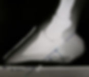

Figure 10. (A) This soft exposure has farrier interest; it clearly defines soft tissue parameters as well as soft tissue lesions. Note opaque lesions proximal to the navicular bone. Palmar angle of navicular bone can be measured from this view as well.
(B) This 65 degree DP was taken with a very soft exposure, clearly defining the thin margin and multiple cystic lesions.
Selecting Exposure Settings
For any radiographic view, the ideal exposure setting will depend on the equipment used (x-ray machine, screens and film, processor, etc.), hoof mass, and the structure of primary interest. I use the terms soft, medium, and hard to describe the exposure settings I select for a particular view, depending on which tissue I am most interested in evaluating. (Note: Specific values for kVp and mAs will depend on the equipment used and the size of the foot being examined, so it is not possible to provide even general guidelines here.)
Soft exposures are for nonbony tissues, such as the horn and corium of the hoof wall and sole, and for the palmar margin of PIII (Fig. 10A). A good soft exposure can reveal differences in radiodensity within the hoof wall which allows differentiation between the laminar corium and the keratinized layers of the hoof wall. When looking for abnormalities at the palmar margin of PIII on the 65 degree dorsopalmar (DP) view, a very soft exposure is needed. If the perimeter of PIII cannot be seen without the use of a hot light, the view should be retaken at an even lower mAs (Fig. 10B).
Medium and hard exposures are used when the structure of interest is bone. Medium exposure is used for bony structures of moderate density or thickness, such as the body of PIII, and for articular surfaces. Hard exposure is used for denser bone or superimposed structures, in particular the navicular bone. I always use a 6:1 parallel grid when using a hard exposure setting.
As this approach illustrates, it is important to tailor the settings to the goal of the examination-to the structure you are most interested in evaluating. "Underexposed" is a relative term. What may seem grossly underexposed to others may be the perfect exposure to show soft tissue detail within the hoof wall or sole, or the palmar margin of PIII.
Unless taking radiographs simply to guide farriery decisions, I take at least two exposures for each view: one soft and one bone detail (medium or hard) exposure. Soft tissue detail is essential, as the nonbony structures surrounding PIII are an integral part of virtually every foot problem.
Factors Affecting Image Quality The diagnostic value of any radiographic examination is determined by the capability of two basic factors: the equipment and the examiner. The skills and knowledge of the examiner are as important as the choice and maintenance of the equipment (x-ray machine, cassettes, screens, film, developing and marking systems, positioning blocks). Not only does the examiner need a good working knowledge of clinical and radiographic anatomy of the foot (including an understanding of the range of normal), s/he needs good radiography skills, from a basic grasp of the geometry of radiation to experience with taking routine radiographic views. Good horsemanship skills are also important.
A disciplined, methodical protocol, designed to provide as much information as possible, is of primary importance if one is to get the most diagnostic value from radiographic examinations. Techniques for ensuring high quality radiographs of the equine foot are described in detail elsewhere1 and will only be summarized here.
Several different factors can affect image quality, and thus limit the amount of accurate information you can obtain from your films:
Preparation of the foot-thoroughly clean the foot of all debris, paying particular attention to the frog sulci.
Beam positioning-the focal area of the primary beam is a zone 4-cm in diameter in the center of the beam; using the light guide or laser pointer, focus the beam on the area of primary interest.
Beam-film relationship-make sure the cassette is perpendicular to the beam on all views; image distortion occurs whenever the film is not perpendicular to the beam.
Subject-film distance-aim for a zero subject-film distance (i.e. cassette in contact with foot) to minimize magnification.
Source-image distance (SID)-use a consistent SID; measure the distance each time, rather than 'eyeballing' it.
Hoof mass-always take into consideration the size of the foot; make separate technique charts for different sized feet, from foal to draft horse.
Radiopaque markers-use markers to clearly and accurately delineate the dorsal hoof wall (lateral views) and, if the horse is unshod, the ground surface (lateral and DP views).
Screens and film-use appropriate film-screen combinations, and replace defective screens and cassettes.
Developing solution-use developer at a consistent temperature set for your technique chart (contrast varies by approximately 10% for every 1 degree F difference); replace weak developing solution.
Film marking-mark each film clearly and accurately; it is a permanent record and your "signature" to colleagues and clients.
Points 3 and 4. Beam-subject-film positioning is every bit as important as the exposure settings used. No matter how good the contrast and detail on the film, the radiograph may be noninformative or misleading if the structures of interest are distorted or obscured because of poor positioning. To minimize image distortion, the cassette must be perpendicular to the beam. Distortion will occur whenever the beam does not strike the film perpendicularly. To minimize magnification, the cassette must be in contact with the foot (i.e. zero subject-film distance).
Point 5. Several authors recommend a SID of 40 in. in order to minimize image magnification. I use a SID of only 24 in. for all radiographs. I have found that the amount of image magnification is negligible between these two SIDs (40 in. and 24 in.), and by using the shorter SID I can use lower exposure settings, thus maximizing the longevity of my x-ray machine and minimizing radiation exposure of personnel. In my experience, beam-subject-film positioning is much more important than the length of the SID in minimizing magnification and image distortion.
Point 7. Let me reiterate how important it is to accurately delineate the dorsal hoof wall with radiopaque material for every lateral foot film. Even at a very soft exposure, you cannot know exactly where the outer surface of the hoof wall is, so you cannot accurately measure dorsal H-L zone width unless the surface of the wall is marked. To paraphrase Murphy's Law, it's on the cases you don't think you'll need it that you'll most wish you'd done it!
Furthermore, having the surface of the hoof wall outlined on every lateral film you take will soon train your eye to recognize subtle increases in dorsal H-L zone width even before you get out your ruler. Almost without thinking about it, you'll have added significantly to your range-of-normal data bank for this particular soft tissue zone.
The Shoe It is not always necessary to remove the horse's shoe for radiographic examination of the foot. Whether or not to remove the shoe depends on the purpose of the examination. In fact, taking routine lateral and DP views with the shoe on can provide valuable information regarding the current shoeing strategy, such as balance, breakover, and loading. However, leaving the shoe on limits the extent of the physical examination.
Depending on the shape, style, and fit of the shoe and horn growth since application, the shoe may obscure certain structures of interest on 45 degree and 65 degree DP views and 65 degree DP-45 degree lateromedial oblique views. The shoe may be superimposed over the palmar margin or wings of PIII, the coffin joint, and/or the navicular bone. In addition, scatter of radiation from the shoe can adversely affect image quality. (Fortunately, most of these structures of interest can be accurately imaged using other views, with the shoe on, without superimposition of metal or scatter of radiation.)
Routine Views "Standard" views of the foot have been suggested as a guideline for practitioners. In my experience they are of limited value, except as screening tools. They are inadequate alone for thorough radiographic examination of the foot. That is because beam orientation, positioning, and exposure settings should be selected in each case based on the purpose of the examination-the reason for performing the examination and the radiographic characteristics of the structure of primary interest.
Below are descriptions of the routine views I take: particular orientations and exposures that I use in most radiographic examinations of the foot. But first, it is important to reiterate that the value of the radiographic examination hinges on how well the physical examination was performed. Physical examination is the single most important aspect of examining the equine foot. With very few exceptions, the objective of the radiographic examination is simply to confirm the findings or suspicions of the physical examination. Relying on radiographic findings in place of a thorough physical examination and without consideration of the history carries the risk of misinterpretation and error, which can be costly.
Lateral
When the principal item of interest is PIII in relation to the hoof capsule and the associated soft tissue zones, the beam should be centered 1/2 - 3/4 in. above the bearing surface (i.e. close to the palmar margin of PIII), midway between toe and heel (Fig. 11). To accurately measure sole depth, distal H-L zone width, and palmar angle, the beam must be centered as close to the palmar margin of PIII as possible. These indices cannot be accurately measured when the beam is centered at or near the coronary band.
In most light horse breeds shod with a normal shoe, the palmar margin of PIII is approximately 1/2 - 3/4 in. above the bearing surface of the wall. In Tennessee Walking Horses and other breeds shod with a raised package or with excess length of hoof wall, the beam must be raised accordingly.
Before taking the radiograph, ensure that the beam is horizontal and perpendicular to the sagittal plane of the foot, the cassette is positioned so that the entire foot is included and is centered on the film, and the cassette contacts the foot and is perpendicular to the beam. (Note: Lining up the heel bulbs by eye as a way of orienting the beam will result in a slightly obliqued view if there is even a slight disparity in the heels, as the beam will not be perpendicular to the sagittal plane of the foot).

Figure 11. For the soft tissue low beam view, the positioning block should be of sufficient height to have the center beam strike the hoof horizontally 0.5-0.75 in above the bearing surface of the wall. Using two blocks assures more accurate information concerning balance and facilitates examination of the lame horse that is unable to stand on one block.
If the positioning block is an appropriate height and the x-ray beam is horizontal and centered between the shoe and the palmar margin of PIII, both branches of the shoe will be precisely superimposed (i.e. only one shoe branch is seen). If the foot is balanced lateromedially, both wings of PIII will also be precisely superimposed.
When the shoe branches are superimposed but the wings of PIII are not (i.e. one shoe branch but two wings are seen), it indicates lateromedial imbalance, which can be confirmed on the DP view. (Note: If the palmar margin of PIII is >1 in. [25 mm] above the block, it may be necessary to raise the beam a little to accurately assess lateromedial balance on the lateral view.) If the shoe branches are not superimposed, it indicates a positioning problem, e.g. the beam is not horizontal and/or it is centered too high on the foot. Sole depth, palmar angle, and dorsal H-L zone width cannot be accurately measured on such a film.
The two exposures recommended for this view are soft and medium. The soft exposure is a "farrier-interest" view, as the information it provides can be of great use to farriers, as well as to veterinarians. Provided the dorsal hoof wall is delineated along its entire length with a radiopaque marker, this view allows accurate assessment of sole thickness, cup depth, medial-lateral balance, digital breakover, dimensions and radiodensity of the H-L and C-E zones, and palmar angle. In addition, it reveals the profile of PIII, and even bone detail along the thin palmar margin. I use this view in 100% of cases, as it is a blueprint for all therapeutic trimming and shoeing strategies.
Commonly diagnosed syndromes such as laminitis (with lamellar zone swelling and/or PIII rotation) and white line disease are easily confirmed with this view. Other lesions that may be evident on this view include fractures in the wing of PIII, proliferative bone changes along the dorsal face of PIII, and the osteoclastic results of keratomas and other space-occupying masses within the hoof wall.
Raised lateral
For a lateral view of the navicular bone or coffin joint, the beam should be centered just below the coronary band and a little closer to the heel-i.e. directly over the navicular bone or coffin joint. Other positioning factors are as described above for the lower lateral view. (Note: On raised lateral films, neither the shoe branches nor possibly the wings of PIII will be superimposed.)
The exposures recommended are medium and hard (using a grid). The medium exposure is best for evaluating the coffin joint. The hard exposure allows evaluation of the navicular bone and surrounding area, including the impar space. By providing a good short-axis view of the navicular bone, this orientation and exposure setting can reveal calcification or avulsion fractures associated with the impar ligament or the navicular suspensory ligaments.
Dorsopalmar view When the principal item of interest is PIII in relation to the hoof capsule, the beam should be centered at the toe, 1/2 - 3/4 in. above the bearing surface of the wall. Ensure that the beam is horizontal and parallel with the sagittal plane of the foot, the cassette is positioned so that the entire foot is included and is centered on the film, and the cassette is perpendicular to the beam.
For routine DP views, the cassette is placed behind the foot, as close to the heels as possible, while making sure the cassette remains perpendicular to the beam. This approach produces a somewhat magnified yet relatively undistorted image. The exposure recommended for this view is soft to medium. This exposure allows evaluation of PIII in relation to the hoof capsule, the hoof capsule in relation to the ground, and thus lateromedial balance. It also allows accurate evaluation of sole depth. These films are farrier-interest views.

Figure 12. This DP view was made with the beam centered over the navicular bone, horizontal to the ground, using a hard exposure and 6:1 grid. The DP 45 degree oblique and the flex lateral taken with the same exposure and grid are also complementing views, and likewise, are not limited by the presence of the shoe.

Figure 13. Positioning for the 65 degree DP view. This positioning block ensures that the cassette is perpendicular to the beam, and thus, minimizes image distortion.
Raised DP
The raised DP view is an excellent projection for evaluating the navicular bone. For this view, the beam is raised approximately 2 in. (depending on the size of the foot) so that it is centered over the navicular bone. The beam is horizontal and all other positioning factors are as described for the lower DP view. The exposure recommended is hard, using a grid (Fig. 12).
If a problem involving the coffin joint is suspected, the raised DP view can be taken at a medium exposure. This view and exposure setting may also reveal fractures through the body or wing of PIII, proliferative bony changes along the palmar margin of PIII, side bone, extensor process lesions (e.g. cysts), and lytic lesions associated with PIII sepsis.
65 Degree Dorsopalmar View
This view is the one most commonly used by clinicians to evaluate the distal margin of PIII and the navicular bone. Many practitioners set up for the 65 degree DP view by placing the cassette in a protective sleeve (tunnel) on the ground, having the horse stand on the tunnel, and angling the beam approximately 65 degree to the ground/cassette. While it is convenient, this practice results in significant distortion of the image, as the beam does not strike the film perpendicularly.
Distorted images of the navicular bone carry an inherent risk of misinterpretation, as they do not accurately represent the architecture of the bone. For example, the normal radiolucent areas within the medullary cavity may appear elongated, widened, or otherwise misshapen on a distorted 65 degree DP.
14A

Figure 14. (A) This radiograph was taken with the bone placed in the positioning block shown in Figure 13 (65 degree DP perpendicular beam to film relationship).
14B

(B) This radiograph was taken with conventional 65 degree DP tunnel projection. Note the measurable distortion that occurs when the beam is less than perpendicular to the film.
It is far better to support the foot on a positioning stand which allows the cassette to be placed perpendicular to the ground and thus to the horizontal x-ray beam (Fig. 13). While some amount of magnification is inevitable, this method ensures that the cassette is perpendicular to the beam, so image distortion is minimal (Fig. 14).
Another potential source of error is failure to adjust for the angle of the navicular bone relative to the ground surface of the foot. Depending on the horse's conformation and on how the 65 degree DP view is taken (e.g. foot loaded or unloaded), the navicular bone may be more upright or more tilted back than expected, which will affect its appearance on the 65 degree DP image. It is worth checking the navicular bone angle on a lateral view (with the foot in position for the 65 degree DP) before taking this view, as some adjustment in hoof position may be needed to get a true dorsopalmar view of the navicular bone. For example, it may be necessary to take a 45 degree DP, rather than a 65 degree DP, to accurately image the navicular bone.
It is possible to get reasonably good detail on 45 degree and 65 degree DP views without removing the shoe, despite the scatter of radiation from the shoe. Often, however, the shod foot cannot be adequately cleaned and the branches of the shoe partially obscure the navicular bone and the wings of PIII. Therefore, it is always best to remove the shoe for these views. For evaluation of the navicular bone, the beam is centered over the navicular area and a hard exposure is taken using a grid. A medium exposure allows evaluation of the coffin joint and the body and wings of PIII. A very soft exposure is indicated for identifying fractures at the distal margin of PIII (solar margin fractures) or soft tissue changes in the toe region. Remember that the bone at the distal margin of PIII is very thin and fenestrated with numerous blood vessels, and the mass of hoof the beam must pass through at this level is relatively small, so a very soft exposure is needed to properly evaluate this area.
Note - For 45 degree and 65 degree DP views, it is very important to clean the foot and distal pastern thoroughly, paying special attention to the heels and the frog sulci, to prevent superimposition of debris over the navicular bone and coffin joint. Even with a well-positioned, high-detail film, failure to thoroughly clean the foot of all debris makes it difficult to properly evaluate these structures.
In addition to thoroughly cleaning out any deep recesses in the frog, I trim the margins of narrow, irregular frog sulci, especially when the frog is very hard and dry. These narrow crevices create an air density over PIII and the navicular bone on 45 degree and 65 degree DP views that can be mistaken for a fracture line.
Packing the frog sulci and bar area with Play Doh or similar material is a common practice when taking 65 degree DP views. I do not pack the foot with anything, as the packing material creates a subtle shadow on the film which interferes with my interpretation of the underlying area. I much prefer the greater detail of an unpacked foot. (To test this yourself, pack only one half of the foot, take a 65 degree DP, and compare the detail between the packed and unpacked sides of the foot.) Another reason I do not pack the foot is because the farrier in me wants to see the outline of the frog and its sulcus-features I am already familiar with from having examined the foot thoroughly before taking radiographs.
65 Degree DP-45 Degree Lateromedial Oblique
When taking 65 degree DP-45 degree lateromedial oblique views, beam position may depend on the structure of primary interest. For example, the beam is centered a little higher for the navicular bone than for lesions in the toe region. Use a soft exposure for the wing of PIII (Fig. 9). This exposure can also be used to evaluate horn and soft tissue lesions in the quarter area, such as quarter cracks, bar cracks, gas-producing abscesses, and puncture wounds.
A medium exposure is suitable for evaluation of the articular margins of the coffin joint. Use a hard exposure (with grid) to evaluate the wing of the navicular bone. This exposure also allows good visualization of the medial or lateral margin of the impar ligament attachment. Calcified lesions within the navicular ligaments, bone spurs, and medullary and cortical changes are all clearly demonstrated on this view.
Flexed Lateral
The positioning block I use for 65 degree DP views can also be used to take a flexed lateral. This view is useful in extremely lame horses that cannot bear weight on the foot. It is also useful for evaluating the coffin joint and navicular area in other horses, as flexing the digit opens the dorsal and palmar aspects of the joint. The shoe does not limit the information gained from this view unless is poses a positioning problem.
Navicular Views Detailed discussion of the navicular bone and associated structures is beyond the scope of this paper. However, some general comments are in order. Perhaps most important is that no one view is adequate for proper examination of the navicular structures. Several views are required to adequately evaluate this part of the foot. In my experience, not only is the 65 degree DP inadequate on its own, it is perhaps the least informative view and the one most prone to misinterpretation, for the reasons discussed above.
I've found the raised DP view, using a hard exposure (with grid), to be a very informative navicular view, provided the angle of the navicular bone is taken into account. This view can reveal abnormal radiolucencies involving the cortex and/or medullary cavity. A 45 degree DP (hard, with grid) complements the raised DP by allowing better evaluation of the wings of the navicular bone. The flexor surface, distal margin (impar ligament attachment), and proximal margin can be evaluated on a raised lateral or flexed lateral view, again taken at a hard exposure with a grid. Oblique DP views can also be useful for revealing navicular fractures. The skyline (palmar proximal-to-distal) view has been advocated by some authors as the view to best image the flexor surface of the navicular bone. It can be a very useful view when taken correctly, as it can show cortical lesions along the flexor surface, particularly within the sagittal ridge. (Fig. 15) It also allows evaluation of the medullary cavity and the relative thickness and density of both cortex and medullary cavity, which can change with the stage of navicular syndrome. In addition, lesions within the deep digital flexor tendon as it runs over the navicular bone may be apparent on this view, particularly if the lesion is calcified.
However, as with the 65 degree DP view, the skyline is easily misinterpreted if the image is distorted by poor beam-film positioning (i.e. when the beam does not strike the film perpendicularly) and if the angle of the navicular bone is not taken into account. Unless the angle of the beam precisely matches the orientation of the navicular bone, it is not the flexor surface that is brought into relief, but the proximal or distal palmar margin of the navicular bone.

Figure 15. This radiograph was taken with the foot placed on a Redden Skyline positioning block that provides perpendicular beam to film relationship. Note that the cassette is seen in the lower left corner. The opaque line crossing the foot is a metal pointer that is set to the palmar angle of the navicular bone.
The pointer aligns the beam, assuring tendon surface relief.
To better understand this concept, take a navicular bone or a similarly shaped object in your fingers and sight down the flexor surface from proximal to distal. Rotate the bone around its long axis (i.e. replicate a change in angle of the navicular bone) and see what difference even a slight amount of rotation makes to the area that is thrown into relief. 16A
Figure 16. (A) Skyline view taken with the beam at pre-determined angle of 41 degree and the cassette positioned perpendicular to the beam. Using a standard setup, the tendon surface is consistently in relief, and elongation is avoided.
16B
(B) Same foot. Skyline tunnel view also taken at 41 degree. Despite proper beam alignment, the tendon surface is superimposed over the body of the bone, and the image is distorted because of elongation.
With very few exceptions, only in grade 2-4 (moderate to severe) club feet will the skyline view, taken as advocated by most authors, show the true flexor surface of the navicular bone. A good way of knowing whether your skyline image is truly showing the flexor surface is to take a series of skyline radiographs of an isolated navicular bone, each at a slightly different proximal-to-distal angle. The view that was taken with the true flexor surface in relief then serves as your benchmark when evaluating skyline radiographs of a horse's foot (Fig. 16A, 16B). Due to the complexity of the foot every effort should be made to minimize distortion.
Note - When a lesion is found in any part of the foot, on any view, bear in mind that it takes two views, one perpendicular to the other (and both taken with the film perpendicular to the beam), to confirm the presence of the lesion and determine its precise location. Measuring the tendon surface angle of the navicular bone (lateral view) as it relates to the ground surface defines the proper beam angle for this view. Using that angle and a positioning block that allows perpendicular beam film alignment assures tendon surface relief.
Digitized Radiography
Digitized radiography (i.e. generation of digital radiographic images) is increasingly being used in equine practice. Advantages include the ability to manipulate the image for enhanced detail (including soft tissue detail) and the ease with which images can be stored and transmitted electronically.
The initial cost is perhaps the greatest disadvantage. The ability to manipulate the image is also a potential disadvantage, as it may result in artifactual loss of detail and thus diagnostically important information. To get the most out of any radiograph of the foot, whether conventional or digitized, it is important to have detailed knowledge of both gross and radiographic anatomy of the foot and an understanding of the range of normal.
And finally, at least one commonly used digital radiography system shrinks the image to 86%. This fact must be borne in mind when taking measurements such as sole depth and H-L zone width from these digitized radiographs.
Venography Venography adds information about vascular perfusion to the details regarding bone and soft tissue structure already available from the plain films (Fig. 17). It can be an extremely valuable addition for diagnostic, therapeutic, and prognostic purposes and for monitoring the response to therapy. Venography can readily be performed in the standing horse, using routine x-ray equipment and easily obtainable supplies [2].

Figure 17. The vascular supply to the digit can be demonstrated with a venogram. A thorough working knowledge of the range of normal variations is essential for accurate assessment.
Key Points
soft tissue pathology is a major cause of foot pain; seek information on the soft tissues in every radiographic examination you perform
develop a methodical approach, and use it every time
select exposure settings for particular views based on the structure of primary interest
develop a series of technique charts that allow for evaluation of different types of tissue (from soft tissue to bone) and different sizes of feet
take at least 2 exposures per view; one for soft tissue detail and one for bone detail
accurately mark the dorsal hoof wall with radiopaque material for all routine lateral views
measure sole depth, dorsal H-L zone width, C-E distance, and palmar angle on routine lateral views for all feet you radiograph to expand your understanding of normal
before taking any films, thoroughly clean the foot of all debris, paying particular attention to the frog sulci
focus the beam on the area of primary interest
make sure the cassette is perpendicular to the beam, on every view, every time
aim for a zero subject-film distance on all possible viewsuse a consistent source-image distance
make sure equipment and developing system are functioning optimally
mark all films clearly and accuratelyinterpret all radiographic findings in light of the history and physical findings
4. Altering Mechanics as a Diagnostic Tool
Using a shoe that alters the mechanics of the foot can be a valuable diagnostic tool during a lameness exam. In addition, the horse's response to the shoe provides valuable insight into how the healing environment within the hoof might be enhanced by altering the mechanics of the foot. This novel approach to examining and treating painful feet is very effective in the majority of footsore horses. Using a mechanical shoe to significantly relieve tension in the deep digital flexor tendon and on all related stress points often improves soundness within minutes of application.
Arriving at a tentative diagnosis after the physical exam, I usually take two survey radiographs with the shoes on and the horse alert (i.e. unsedated). The routine lateral and DP views described above provide all the information needed to assess hoof mass and balance, and to identify the majority of common soft tissue lesions.
Most lame horses do not have bone pathology, so the solution in these cases generally relies on an understanding of how subtle changes in soft tissue parameters affect the overall health and soundness of the foot. The primary problem often involves soft tissue compression and associated vascular compromise which, in many cases, is the underlying cause of the pain and deteriorating hoof mass.
I grade the mechanical effect of the shoe or other therapeutic device as follows: one point is given for every 2 degrees increase in palmar angle (with the horse bearing weight on the limb). Thus, a shoe with a mechanical score of 1 raises the palmar angle by 2 degrees; this is a "low-mechanics" shoe. A device with a mechanical score of 5 is one that raises the palmar angle by 10 degrees; the mechanical effect is described as moderate or intermediate. Raising the palmar angle 20 degrees (such as is done for horses with acute laminitis) has a mechanical score of 10; this is a "high-mechanics" device.
Depending on the degree of lameness, the tentative diagnosis, and the horse's training schedule, I usually start with a low-mechanics shoe. I move up the scale as needed, guided by the horse's response and how readily the horse can unload the painful area in the particular shoe. I observe the horse at a walk and a trot, on soft and hard footing, on a slope, and on a 30-ft diameter circle. Figures 18A and 18B illustrate the effects of a high-mechanics shoe on palmar angle and functional breakover.
18A
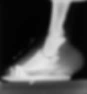
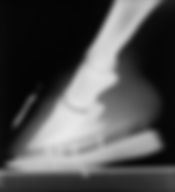
Figure 18. (A) Before and after application of a four-point rocker rail. This was apparently caused by a large cystic lesion involving the navicular bone. This shoe was used to define and treat heel pain. See DP view of Figure 12.
18B

(B) This shoe was used to treat a Thoroughbred race filly presented with heel pain that was caused by severe caudal rotation (negative palmar angle). Note: Capsular palmar angle A and palmar angle B created with the ground surface.
5. Concluding Remarks
Effective examination of the foot hinges on an appreciation of its normal structure and function, encompassing the hoof capsule, soft tissues, vasculature, and bone. Clinical and radiographic examinations are merely discovery exercises, aimed at identifying the area(s) in which structural or functional integrity has been lost. This aim cannot be achieved without an understanding of normal, including an appreciation for the sometimes wide range of normal dictated by the horse's breed, age, environment, and use.
It is not enough for us to reach a medical diagnosis; our examinations must have the dual goal of directing us toward a solution to the horse's problem, both immediate relief and a long-range plan for restoring and preserving structural and functional integrity. That goal can be met only when our examinations are aimed at collecting as much specific information as possible, about every component of the digital unit.
Although it is important to tailor specific techniques to the goals of the examination, it is equally important to develop and practice a disciplined, methodical approach to both clinical and radiographic examinations. Errors of omission and misinterpretation are minimized when the examiner collects as much information as can be gained from both procedures and considers the significance of the findings in total.
Perhaps the single most important addition practitioners can make to their examination procedure is a radiographic protocol which includes views and exposures that provide detailed information about the soft tissues of the foot. Making it part of every radiographic examination will greatly enhance your diagnostic capabilities by allowing you to develop an eye for fine detail and thus subtle abnormalities that would otherwise have been missed.
References and Footnotes
1. Redden RF.Radiography of the equine foot-techniques for enhancing the quality of your films.Versailles: Nanric Inc., 2002; 1-24.
2. Redden RF. A technique for performing digital venography in the standing horse.Equine Vet Educ2001;3:172-178.
a Intropaste, Barium Sulfate Paste, Lafayette Pharmaceuticals, Inc., Lafayette, IN 47904 or Yorba Linda, CA 92887.
b Plexigas, Rohm GmbH & Co., KG, D-64293 Darmstadt, Germany.
AAEP PROCEEDINGS / Vol. 49 / 2003 Pages 169-185